Jun 5, 2019
Restoring Vision with Stem Cells
Dr. David Gamm is a pediatric ophthalmologist investigating how stem cells can be used as a potential treatment for retinitis pigmentosa (RP) and age-related macular degeneration (AMD). Dr. Gamm’s research is supported through Fighting Blindness Canada’s Restore Vision 20/20 Initiative. To learn more about Dr. Gamm’s FBC-funded research, please read this story about his efforts to develop new sight-saving treatments. This is an account of his presentation at Fighting Blindness Canada’s Vision Quest Conference in Vancouver, April 27, 2019.
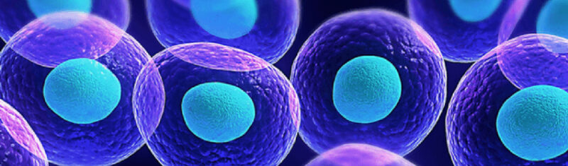
What is a Stem Cell?
If a healthy cell becomes diseased it will usually be able to heal itself, either through the actions of the immune system or by treatment with a therapeutic, such as antibiotics. But some diseases are degenerative, meaning that instead of healing itself, the cell will break apart and die. Once these cells are dead we cannot put them back together.
Some tissues, like the skin, liver, gut lining and blood, have special “hidden” cells in them called tissue-specific stem cells. These cells lie dormant until an injury or disease occurs. They are then induced to make more of themselves and repopulate any cells that have been damaged or destroyed.
Unfortunately, the retina does not have the capacity to repair itself once cells have been destroyed. This is why there are no cures for diseases such as age-related macular degeneration (AMD) and retinitis pigmentosa (RP) – the body can’t replace these parts once they have been destroyed.
Once degeneration occurs in these tissues we have limited options for treatment. The two main courses of treatment are to bypass the cells by getting other cells to do the job for them (prosthetic chips, optogenetics) or we can replace these tissues with stem cells. Research is focusing on two main cell types for replacement: photoreceptors and retinal pigment epithelium (RPE). There are other cell types that can potentially be replaced but these present a significant challenge.
Diseases of the Retina:
The retina is like the film in a camera – it covers the back of the eye and is responsible for capturing the light that will make the image your brain sees. The retina has many layers of different kinds of cell types. These lie in the retina in a specific order. The deepest layer of the retina contains two very important cell types.
- Photoreceptors (rods and cones) receive light coming into the eye. They pass this signal through the retina to the ganglion cells and then to the brain, which “sees” the image. If these cells are damaged directly it will cause an eye condition called retinitis pigmentosa (RP).
- Retinal Pigment Epithelium (RPEs) are helper cells. They make sure that the photoreceptors are working properly. When these cells degenerate, it causes the photoreceptors to die a secondary death and vision loss occurs. This is what happens when someone is affected by macular degeneration.
Another type of potential treatment that is being widely talked about is gene therapy. In this case the disease must be specific to one particular gene that can be targeted. However, not all degenerative diseases are caused by one particular gene. The hope is that a stem cell treatment could be applied to a much broader group, including diseases that cannot be treated by gene therapy.
More importantly, the stage of degeneration is critical to determining if someone is a good candidate for stem cells. If the disease is caught early on, the focus should be on protecting the remaining cells, not on replacement. If it is too late, the entire retina may be damaged; therefore, a few stem cells will not make a significant difference. This window for replacement must be taken into account when considering if someone might be a suitable candidate for a stem cell treatment.
Sources of Retinal “Spare Parts”
Stem cell therapy consists of two main parts: the manufacture of “spare parts” (stem cells) and the installation of these cells.
There are different types of cells that scientists are studying to develop new therapies. The research in Dr. Gamm’s lab is focused on a kind of cell called induced pluripotent stem cells (iPSCs). These specialized cells were first generated in 2006-2007 when scientists discovered how to take adult cells (such as skin cells) and reprogram to behave like embryonic stem cells, which have the potential to make all the different cell types in the human body! These iPSCs can be used to help study disease, but also have the potential to be put into patients to replace lost cells.
There are two key advantages to using iPSCs: 1) they are an authentic source of retinal cells, and 2) they are in unlimited supply.
Making Retinal “Spare Parts”: Retinal Pigment Epithelium (RPE)
Induced pluripotent stem cells (iPSCs) have the potential to become any of the cells in your body. Scientists use various techniques to coerce the cells to go down a specific path and become the cell types they want. To do this in the lab, researchers use various chemicals, physical forces and other cellular cues, all while making sure the finished cells will be safe for use in humans.
When retinal pigment epithelial (RPE) cells are made, they are brownish-black cells that look like little stop signs. They form into a nice sheet and can perform all the functions that normal human RPE cells can do. Making RPE has several advantages. Firstly, it is one of the easiest cells to produce, isolate and culture from stem cells. Because of this, all current clinical trials are using RPEs made from stem cells. Another advantage is that these cells do not need to “fit in” to other cells or make a connection. This makes it easy to “install” them in a patient’s eye.
Making Retinal “Spare Parts”: Photoreceptors
Dr. Gamm’s lab is working with photoreceptor cells. These are the more challenging of the cell types for several reasons:
- Photoreceptors require connectivity to other cells. Not only do they interact with RPEs, they also need to make a “handshake” with other retinal cells to pass along their signals to the brain.
- Photoreceptors do not grow in isolation. Instead they must grow in a community of cells that will ultimately make up the entire retina and all its complex layers. Once these cells mature, the photoreceptor cells need to be extracted for therapeutic use.
After many years of work, Dr. Gamm and his team now have a “product” that looks like a viable replacement part – the photoreceptors have all the features of rods and cones, they can detect photons of light, and they can detect light at the appropriate wavelengths. But at the moment, these promising developments are confined to petri dishes. Their next step is to evaluate whether these photoreceptors can respond to light in living organisms, and then go through the entire process carefully to make sure that these cells would be safe for human use.
So how do these spare parts get installed in the correct location in the eye? This is the question that Fighting Blindness Canada is funding Dr. Gamm’s team to study.
Donor Cell Installation
Clinical trials with retinal pigmented epithelial (RPE) cells have already started to tackle the problem of how to insert these cells. The simplest way to get under the retina is to form a small hole and lift it up so that the cells can be injected in. This method will inevitably evolve over time as new research is done into the most effective and safe way to deliver cells.
Another important consideration is that it would be beneficial not just to randomly inject the cells into the space evacuated by the disease, but instead, to make sure that the cells are oriented in a specific direction. This can be accomplished by growing the cells on small scaffolds that can be placed in the eye.
Learning how to install these cells remains one of the biggest challenges to making this treatment a reality! Fighting Blindness Canada is proud to be funding Dr. Gamm’s essential research that aims to overcome this hurdle.
Whose Stem Cells Do We Use?
There are several potential ways to make stem cells for retinal research.
- The technology exists to make personalized cells for every individual – however this is very expensive and therefore is not likely a viable option.
- “One-Size-Fits-All” cells would, in theory, not be recognized and rejected by our immune system.
- A bank of partially matched cells could be developed and would be similar to the banks that are currently used for organ transplantation. These would not be perfect matches, but much closer than a random cell type.
- A “universal cell” that can evade the immune system.
Update on Stem-Cell Clinical Trials
There are several ongoing clinical trials that are testing retinal pigment epithelial (RPE) cell replacement therapies on human subjects. Early trials were done by delivering unstructured RPEs behind the retina. The results showed that these procedures were safe – there were no tumours or ill effects – but there was also no benefit to patients. Later, different research groups started growing RPEs onto fabricated sheets and implanting these grafts in late-stage AMD patients. We reported on the promising results from one of these trials in this story.
These early trials showed that this potential treatment was safe and seemed to slow down the progression of the disease. However, because it was not replacing the damaged photoreceptor cells, vision could not be restored.
Dr. Gamm’s research has been the recipient of the FBC Restore Vision 20/20 grant. This is allowing them to do critical experiments that will bring them closer to their goal of starting a clinical trial with photoreceptor cells in the next 2-3 years.
Future Expectations
Currently, there are no approved stem cell therapies. It is critical to remember that not all stem cells are created equally, and many companies are using sham stem cell claims to make a profit. Be wary of stem cell clinics that don’t fully explain the products they are implementing, charge fees, promote themselves using patient testimonials, or make far reaching claims.
While significant progress has been made, it is important to look to the future with realistic expectations. Stem cell research holds tremendous promise, but it is important to recognize that the earliest stem cell treatments will not reverse all vision loss, nor will they be a “cure” for everything. It is going to take years of dedication and multiple clinical trials to have a meaningful impact on people’s vision. It is hoped and anticipated that stem-cell based therapies will have a place among a variety of treatment options.
To learn more about stem cell therapies for vision loss, visit our Must Read Resource on Stem Cell Therapy for Vision Loss.
Join the Fight!
Learn how your support is helping to bring a future without blindness into focus! Be the first to learn about the latest breakthroughs in vision research and events in your community by subscribing to our e-newsletter that lands in inboxes the beginning of each month.